Cryo Transmission Electron Microscope
Cryo Transmission Electron Microscope
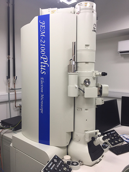
Our cryo-Transmission Electron Microscope (cryo-TEM), model JEM-2100Plus: made and installed by JEOL UK Ltd. The microscope is capable of imaging flash-frozen (with liquid nitrogen (LN2)) specimens at accelerating voltages up to 200 kV.
Its auto focus/contrast/brightness/exposure/montage, drift compensation and stage navigation features revealing observed details as small as 0.25 nanometres, or several hundred thousand times thinner than a human hair, while operating at a typical magnification in excess of 50,000x.
Gatan model 626 cryoholder
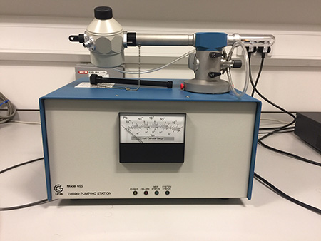
Gatan model 626 cryoholder sitting, inserted, a-top its model 655 vacuum pumping station. The cryoholder must be pumped down overnight to high vacuum, e.g. 10^6 Torr, in order to ensure its thermal stability remains as stable as possible when its end (spherical) chamber is cooled with liquid nitrogen (LN2) down to 196°C, typically operating in the range -180 to 184°C i.e. the specimen under observation will not drift during imaging (which would then blur the CCD output, and resolution lost).
When the cryoholder has been under vacuum for several hours and sufficiently desiccated, it is cooled in a separate cryo-station, again down to LN2 temperatures, to then be rapidly inserted rapidly, within a minute, into the goniometer of the JEM-2100Plus above.
Polaron E7000 by BioRad
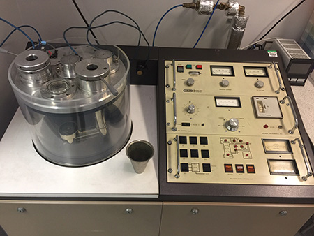
The well revered Polaron E7000 by BioRad, which is used for freeze fracture. Here, frozen specimens, often with liquid nitrogen or possibly liquid ethane, such as tissue or membrane samples (even emulsions, gels or liquids) may be fractured (or cracked) across their weakest point to reveal a plane of embedded structural detail e.g. the position and orientation of proteins.
This can prove potent in understanding the underlying ultrastructure present, for functional studies, especially if +/- disease states are compared.
Ultracut E Ultramicrotome
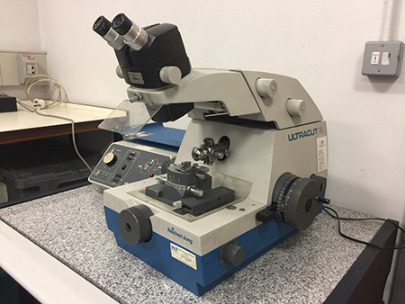
Reichert-Jung made a venerable Ultracut E Ultramicrotome, which is ideal for us to obtain ultra-thin, micron-thick specimens (a human hair is typically 25 to 50 microns thick), using both diamond and glass knives to thinly slice resin-embedded samples. The samples are now thin enough for the electron beam in a TEM to pass through, usually with added contrast possible in the image by negatively staining or metal-coating (with a thin sputter) it prior to insertion into the microscope.
easiGlow automated glow discharge unit
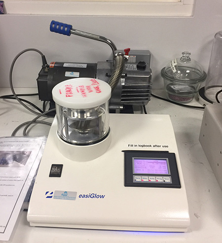
easiGlow automated glow discharge unit by Ted Pella, Inc. with, at rear, its vacuum pump.
For rapid cleaning of surfaces or for introducing an even charge across the surface of TEM specimen grids that may aid protein molecule adhesion, or orientation, prior to flash-freezing, and/or negatively staining, samples before single particle or tomographic analyses.
JEOL model JEM-1230 TEM

The JEOL model JEM-1230 TEM, housed within QMUL’s Nanovision centre, for imaging with accelerating voltages up to 120 kV. A side-entry non-cooled Morada 2K camera is seen, coloured silver, at the bottom left hand side of the main column, which is ideal for ascertaining the quality of biological samples, perhaps prior to going forward to cryo-work with the 2100Plus.
However, it is used daily to produce very nice images of ultra-thin tissues samples, ~18,000x magnification, and negatively stained single protein molecules/particles ~20 nm in diameter, with a resolution of 1.7 nm in projection, have been observed with a narrow field of view (at 80,000x max.).